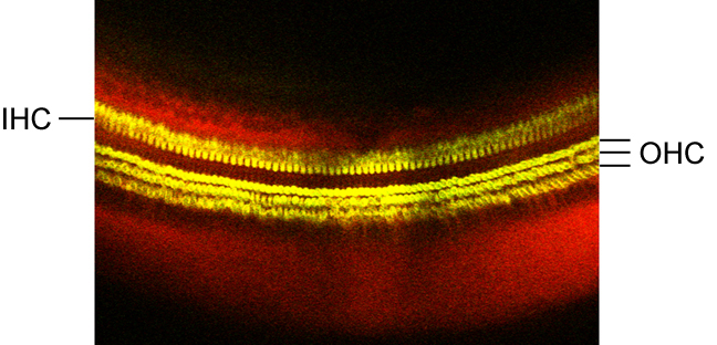
The organ of Corti
A
view looking down onto the organ of Corti from the direction of the
stereocilia.
The cells are stained in yellow with a membrane dye, FM 1-43. The
surrounding fluid
is stained in red with the dye Dextran Oregon Green 514. The three rows
of outer
hair cells (OHC) are seen in the lower part, and the row of
inner hair cells (IHC) in the upper part of the figure. The picture was
made by
Dr. M. Nowotny using the
two-photon confocal laser-scanning microscope.
With this set-up we are performing Ca2+ imaging experiments,
membrane re-cycling
observations and morphological studies.
© 2005 A. W. Gummer.
![]()